
Radiology Modalities Explained: Understanding Medical Imaging Techniques
From X-rays to MRI and CT scans, understanding different imaging techniques can help patients and radiology stakeholders alike to gain insights into their applications, benefits, and impact on radiology workflow.
Radiology modalities are the different techniques and technologies used to capture images of the inside of the body. These images can provide valuable information to help doctors make accurate diagnoses and develop effective treatment plans.
Understanding these modalities is essential for healthcare professionals and patients alike, as it enables informed decision-making and facilitates effective communication between radiologists, physicians, and patients.
What are Radiology Modalities?
Radiology modalities, or medical imaging modalities, are techniques used to create images of the human body for diagnostic and therapeutic purposes. These modalities utilize different technologies to visualize the internal structures and organs, helping healthcare professionals detect abnormalities, diagnose diseases, and guide treatment plans.
Types of Radiology Modalities
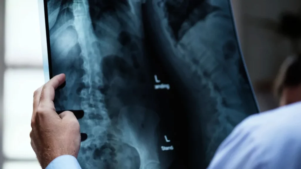
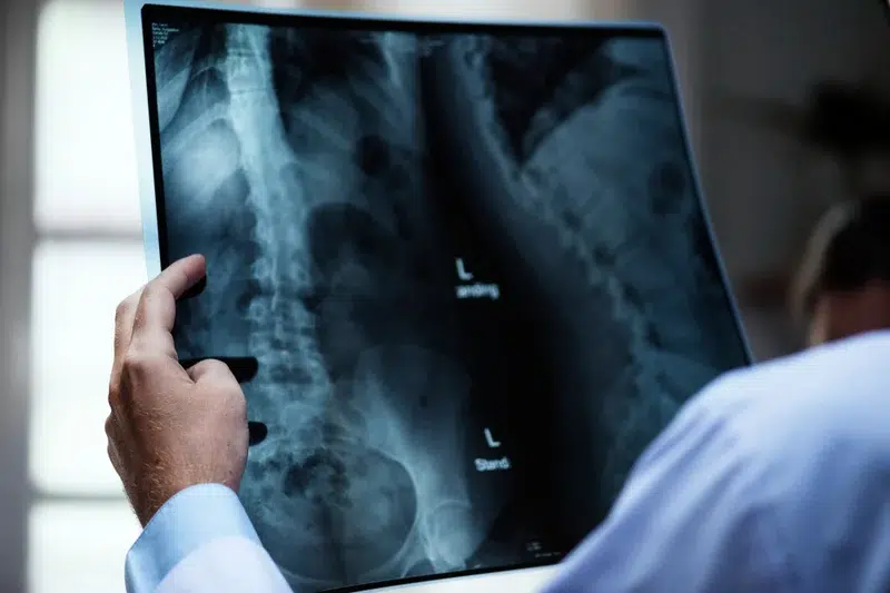
X-rays
X-rays are perhaps the most well-known type of radiology modality, these work by passing radiation through the body, which is then detected on the other side by a film or digital detector. The images produced by X-rays are black and white and show the density of different tissues within the body.
X-rays are commonly used for:
- Fracture diagnosis and assessment
- Lung infection detection
- Dental imaging and evaluation
Duration: this type of modality usually takes between 10 and 15 minutes.
| PROS | CONS |
|
|
Fluoroscopy
Fluoroscopy is a real-time imaging technique that uses X-rays to visualize the body’s internal structures in motion.
Fluoroscopy is often used during surgeries, particularly for digestive, urinary, or reproductive systems procedures. It can also be used to guide the placement of catheters or other medical devices. One of the benefits of fluoroscopy is that it allows doctors to see the movement of organs or structures in real-time, which can help with diagnosis and treatment planning.
However, fluoroscopy does expose patients to radiation, which can be harmful in large amounts. Doctors carefully consider the benefits and risks of all medical imaging modalities before recommending fluoroscopy. They may also take steps to reduce radiation exposure, such as using a lead apron or limiting the imaging duration.
Fluoroscopy is commonly used for:
- Barium enema procedures
- Cardiac catheterization
- Joint injections
Duration: 30 minutes – 2 hours
| PROS | CONS |
|
|
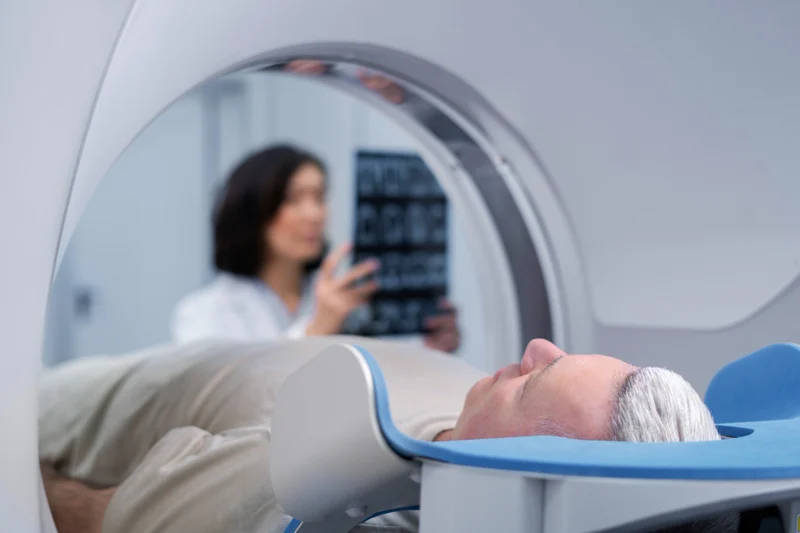
CT Scan (Computed Tomography)
CT, or computed tomography, is a non-invasive diagnostic imaging technique that uses X-rays and computer technology to create cross-sectional images of the body. CT scans are incredibly helpful in detecting internal injuries and diseases, as they can provide detailed, 3D images of organs, bones, and other tissues that traditional X-rays may not capture.
During a CT scan, the patient will lie on a table that moves through a circular opening in the scanner. As the table moves, the scanner takes multiple X-ray images of the body from different angles. The computer then uses these images to create a 3D image of the area being scanned. CT scans can be done with or without contrast dye, which is injected into the patient’s bloodstream to help highlight certain areas of the body.
CT scans are commonly used to diagnose conditions such as cancer, heart disease, and lung disease, as well as to monitor the progression of certain conditions. They are also frequently used in emergency situations to quickly diagnose injuries and determine the extent of internal damage.
While CT scans are generally considered safe, they do expose the patient to radiation, so they should only be done when necessary. Patients who are pregnant or have kidney problems may not be able to have a CT scan with contrast dye.
CT Scans are commonly used for:
- Tumor detection and staging
- Vascular disease evaluation
- Internal injury assessment
Duration: 20-25 minutes
| PROS | CONS |
|
|
MRI (Magnetic Resonance Imaging)
A non-invasive medical imaging technique that uses strong magnetic fields and radio waves to create detailed images of the body’s internal structures. Unlike X-rays or CT scans, MRI does not use ionizing radiation, making it a safer option for patients.
MRI is beneficial for visualizing soft tissues and structures, such as the brain, spinal cord, muscles, and organs. It can detect abnormalities such as tumors, blood clots, and injuries to the soft tissue.
During an MRI, the patient lies down on a table that slides into a large tube-shaped machine. The machine then creates a strong magnetic field around the body, causing the atoms in the body to align with the field. Radio waves are then used to stimulate the atoms and create signals that are detected by the machine and translated into images.
MRI is a versatile tool that can produce different types of images depending on the specific technique used. Some common types of MRI include T1-weighted images, T2-weighted images, and diffusion-weighted images. These different types of images can provide additional information about the tissues being imaged, such as the presence of inflammation or water content.
Despite its benefits, MRI has some limitations. It can be a relatively slow imaging technique, with some exams lasting up to an hour. The machine is also very sensitive to movement, so patients must remain still during the exam. Additionally, the magnetic field can interfere with certain types of medical devices, such as pacemakers or metal implants.
MRI is commonly used for:
- Brain and spinal cord imaging
- Soft tissue evaluation
- Multiple sclerosis diagnosis
Duration: 45 minutes – 1 hour
| PROS | CONS |
|
|
Ultrasound
Also known as sonography, this imaging modality uses high-frequency sound waves to create real-time images of the body’s organs and tissues. It is commonly associated with prenatal imaging, allowing healthcare providers to monitor the growth and development of a fetus. Ultrasound is also used to examine the abdomen, pelvis, heart, and blood vessels.
Ultrasounds are commonly used for:
- Prenatal imaging and monitoring
- Abdominal and pelvic evaluation
- Cardiac and vascular imaging
Duration: 30 minutes – 1 hour
| PROS | CONS |
|
|
![]()
PET Scan (Positron Emission Tomography)
Another commonly used radiology modality that has revolutionized medical imaging techniques. This diagnostic tool uses small amounts of radioactive materials called radiotracers, which are injected into the patient’s body. As these radiotracers are absorbed by the body, they release energy in the form of positrons, which combine with electrons to produce gamma rays. These gamma rays are then detected by the PET scanner, which produces images that are analyzed by radiologists to diagnose various conditions.
PET scans are particularly useful in detecting cancer, as the radiotracers used in the scan tend to accumulate in cancer cells more than normal cells, making it easier to identify tumors. PET scans can also be used to evaluate the extent of cancer, to assess the effectiveness of cancer treatments, and to detect cancer recurrence.
Apart from cancer, PET scans can also be used to diagnose various other medical conditions, such as neurological disorders, heart disease, and other metabolic disorders. PET scans are non-invasive and usually take around 30 minutes to an hour to complete.
Overall, PET is a powerful diagnostic tool that has transformed the way radiologists diagnose and treat various medical conditions. With its ability to detect and diagnose cancer at an early stage, PET has been instrumental in improving patient outcomes and increasing survival rates.
Pet Scans are commonly used for:
- Cancer diagnosis and staging
- Brain function evaluation
- Heart disease assessment
Duration: 1.5 – 2 hours
| PROS | CONS |
|
|
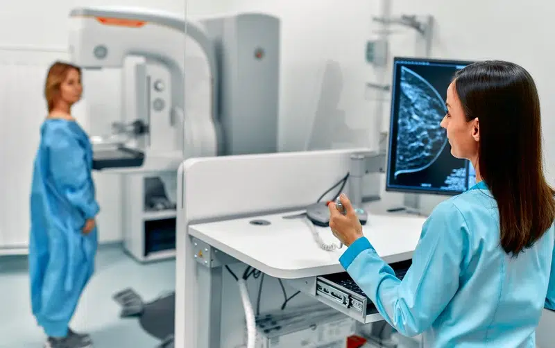
Mammography
A type of radiology modality that is primarily used to detect and diagnose breast cancer. It involves the use of low-dose X-rays to create images of the breast tissue. Mammography is a widely accepted screening method for breast cancer and is recommended for women above the age of 40.
During mammography, the breast is compressed between two plates for a few seconds to get an accurate image. The resulting image is called a mammogram, which can be used to detect any abnormalities, such as lumps or changes in the breast tissue. Early detection of breast cancer is crucial for successful treatment and improved outcomes.
Mammography can be performed in two ways:
- Screening mammography: performed on asymptomatic women to detect any potential abnormalities in the breast tissue.
- Diagnostic mammography: performed on women who have symptoms such as breast lumps or discharge.
One of the challenges of mammography is the high rate of false positives, where a mammogram indicates a potential abnormality that is not cancerous. This can lead to unnecessary biopsies and anxiety for the patient. However, improvements in mammography technology and interpretation have led to a decrease in false positives.
In addition to traditional mammography, there are also other types of mammography, such as tomosynthesis or 3D mammography. Tomosynthesis uses multiple X-ray images to create a 3D image of the breast, which can help to reduce false positives and detect smaller cancers.
Mammography is commonly used for:
- Breast cancer screening
- Detection of breast abnormalities
Duration: 30 minutes
| PROS | CONS |
|
|
Role of Radiology Modalities in Diagnosis
With these imaging techniques, it is easier to identify various medical conditions that are hidden beneath the skin or inside the body. The importance of radiology modalities is highlighted by the fact that doctors rely on them to confirm or rule out diagnosis based on symptoms and other medical tests.
Radiology modalities can help identify many different medical conditions, including bone fractures, cancers, and cardiovascular disease. X-rays and CT scans are particularly useful in identifying fractures and joint injuries. In contrast, MRI and PET scans can provide detailed images of soft tissues, such as organs and blood vessels, which helps identify problems in these areas.
Ultrasound is often used in obstetrics and gynecology to examine the reproductive organs, identify cysts and tumors, and monitor fetal growth during pregnancy.
The use of mammography, an imaging technique that uses low-energy X-rays to examine the breast, has contributed significantly to the early detection of breast cancer, making it easier to treat. By examining the mammography images, radiologists can detect abnormal growths in the breast tissue and distinguish them from non-cancerous growths. In this way, mammography has revolutionized the diagnosis and treatment of breast cancer.
Final thoughts
Understanding the various radiology modalities is essential for both healthcare professionals and patients. Each modality has its unique strengths and limitations, and choosing the most appropriate one will depend heavily on accurate communication.
Imaging modalities and their impact on patient-radiologist communication
A radiology center’s goal is to provide accurate, reliable, and timely imaging results to their patients.
Radiology modalities can be confusing for patients who may need help understanding the technical jargon or the imaging process. Patients should feel empowered to ask questions, express concerns, and provide relevant medical history to assist in interpretation. Along the same line, referring physicians should maintain open and specialized communication with radiologists, discussing clinical indications and sharing pertinent information.
In addition, having a specialized radiology scheduling service that understands the complexities of different imaging procedures helps optimize appointment scheduling and minimizes delays.
What happens when radiology practices and imaging centers stop settling for outdated and general scheduling methods and switch to a radiology call center?
Request a Consultation now and unlock the efficiency and cost-effectiveness your radiology practice deserves.